
Revolutionary SPIFFI Microscopy Technique Captures Super-Resolution Live Cell Videos in a Single Exposure

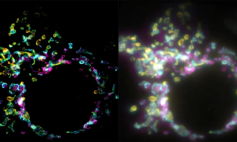
In the relentless pursuit of visualizing the microscopic machinery of life, a team of researchers has unveiled a groundbreaking fluorescence microscopy technique capable of generating high-resolution, sharp imagery from a single exposure. Developed by scientists at the École Polytechnique Fédérale de Lausanne (EPFL), this new method—dubbed SPIFFI, which stands for spatial polarization-induced fluorescence fluctuation imaging—promises to fundamentally change how biologists observe fast-paced intracellular dynamics. Detailed in a recent publication in the prestigious journal Nature, the innovation sidesteps the historical speed limitations of super-resolution microscopy, opening the door to real-time, nanoscale cinematic views of living cells.
The breakthrough, spearheaded by researchers Wei Guo, Lely Feletti, and Aleksandra Radenovic from EPFL’s Laboratory of Nanoscale Biology (LBEN) within the School of Engineering, addresses a fundamental bottleneck in modern cellular biology: the trade-off between spatial resolution and temporal acquisition speed.
The Historical Evolution and Limitations of Super-Resolution
For decades, traditional light microscopy was bounded by the classical diffraction limit of light, a physical constraint first articulated by Ernst Abbe in 1873. This rule dictates that light microscopes cannot resolve details finer than roughly half the wavelength of light used—typically around 200 to 300 nanometers. Anything smaller than this threshold blurs into an indistinguishable smudge.
In the late 20th and early 21st centuries, the advent of super-resolution techniques—such as stimulated emission depletion (STED) microscopy, photoactivated localization microscopy (PALM), and stochastic optical reconstruction microscopy (STORM)—shattered this barrier. These pioneering methods earned their inventors the Nobel Prize in Chemistry in 2014, fundamentally revolutionizing molecular and cellular biology by allowing researchers to image subcellular structures down to a few nanometers.
However, these conventional super-resolution frameworks carry a heavy operational cost: time. To piece together a single, crisp super-resolved image, these methods typically require the capture of hundreds or thousands of sequential individual frames. Each frame captures stochastic fluctuations or targeted states of fluorescent molecules over time, which are then computationally reconstructed into a final, high-resolution composite.
While this multi-frame approach works remarkably well for fixed, static samples mounted on microscope slides, it creates a nearly insurmountable hurdle when applied to living cells. Cellular environments are not static museums; they are bustling, dynamic systems characterized by constant movement, rapid molecular trafficking, and fast-evolving structural changes such as cell division, mitochondrial remodeling, and membrane fusion. Attempting to capture these rapid processes with multi-frame methods often results in severe motion blur or temporal artifacts, rendering the final image inaccurate or completely uninterpretable.
The Mechanics of SPIFFI: Polarization and Single-Frame Clarity
To overcome the temporal limitations of traditional multi-frame approaches, the EPFL research team looked past temporal information and turned instead to spatial polarization.
When fluorescent molecules—often referred to as fluorophores, which are attached to specific cellular proteins or structures—are excited by a light source, they emit fluorescent light. Crucially, the light waves emitted by these molecules do not radiate uniformly in all directions. Instead, they oscillate preferentially along specific axes depending on the precise physical orientation of the individual molecule.
SPIFFI harnesses this natural phenomenon through an innovative optical hardware configuration. The microscope splits the incoming fluorescent light emitted by the sample into four distinct, polarization-sensitive channels simultaneously. By comparing the variations and intensity patterns across these four channels within a single snapshot, the system extracts rich structural details that would otherwise remain hidden in a conventional, non-polarized view.
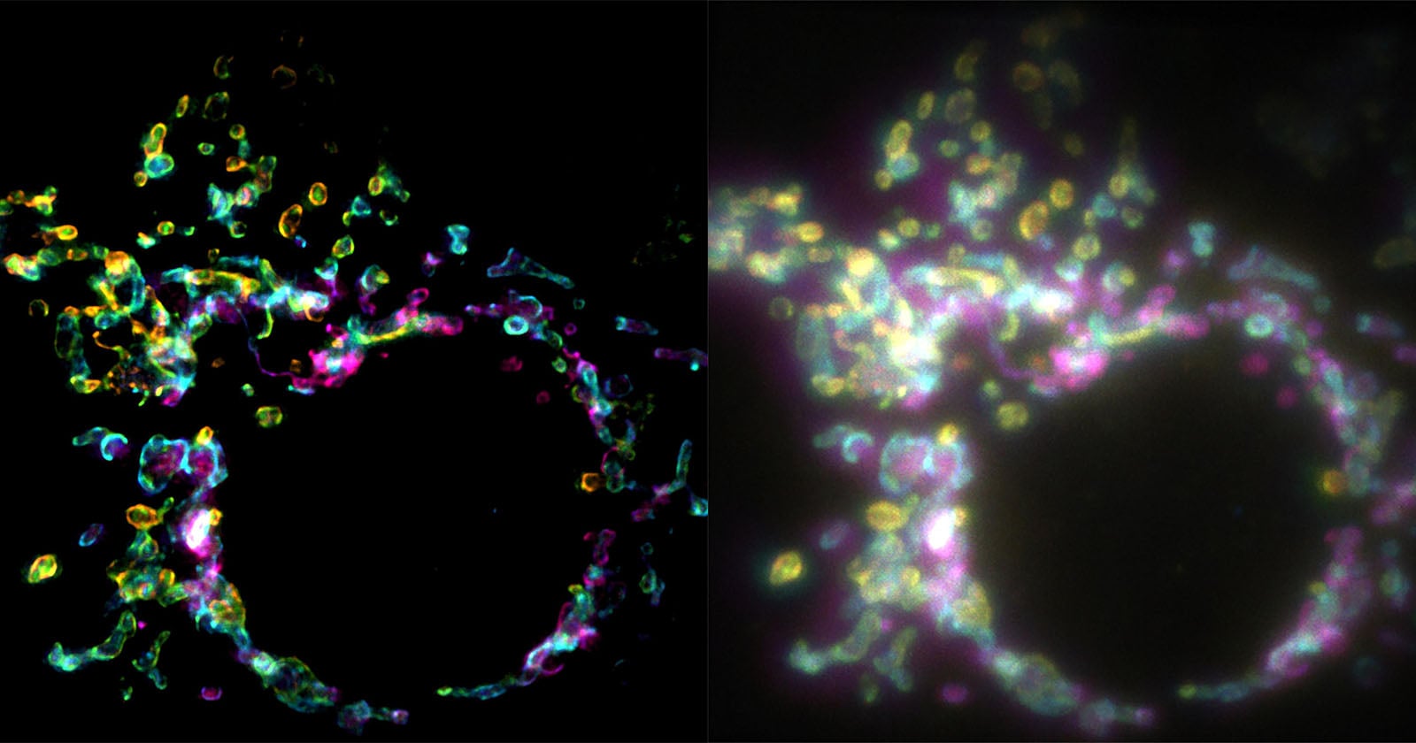
According to Wei Guo, a PhD student at LBEN and the first author of the research paper, this fundamental shift in methodology changes the entire paradigm of advanced microscopy. "Essentially, previous approaches used temporal information to resolve spatial resolution, but this doesn’t work very well on living cells," Guo explained.
By analyzing polarization gradients within a single exposure, SPIFFI bypasses the need to accumulate data over hundreds of frames. "With previous techniques, taking many images would only result in one super-resolved frame," Guo noted. "With SPIFFI, every frame is super-resolved, meaning we can now produce super-resolution videos of live cells."
Experimental Validation and Performance Metrics
During rigorous testing and experimentation, the EPFL team demonstrated that SPIFFI can reliably improve image resolution by a factor of two using just a single image capture. The technique successfully resolved delicate cellular structures measuring approximately 160 to 170 nanometers in size—a significant leap beyond the capabilities of standard widefield fluorescence microscopy.
Furthermore, the research team applied SPIFFI to capture complex, transient biological events that were previously exceedingly difficult to document clearly, such as the outer membrane dynamics of mitochondria during cellular splitting and fusion.
The versatility of the system does not stop at single-frame super-resolution. The researchers successfully integrated SPIFFI’s primary optical output with existing fluctuation-based post-processing algorithms. Through this hybrid approach, the team achieved an even finer effective resolution of approximately 80 nanometers, bridging the gap between hardware innovation and computational enhancement.
Aleksandra Radenovic, head of the Laboratory of Nanoscale Biology, emphasized the broader utility of the technique in a statement regarding the publication. She noted that SPIFFI "can capture fast-moving processes within cells, while enabling high-throughput, multi-dimensional imaging beyond the limits of conventional microscopes."
Practical Implementation and Future Horizons
One of the most promising aspects of the SPIFFI technology is its accessibility and potential for integration into existing laboratory infrastructure. Rather than requiring researchers to invest in entirely new, prohibitively expensive microscopy systems, SPIFFI’s specialized optical hardware is designed to be retrofitted into many existing fluorescence microscopes. This compatibility drastically reduces the barrier to entry for academic labs and commercial research facilities alike.
Building on this success, the EPFL research team is actively working on the next phase of development. Their current engineering objective is to make the SPIFFI optical setup significantly more compact, which will further streamline its usability, improve alignment stability, and facilitate broader adoption across diverse biological disciplines.
Implications for Life Sciences and Medicine
The introduction of single-exposure super-resolution imaging arrives at a critical juncture for biomedical research. As scientists strive to understand the subtle mechanisms driving viral infections, neurological disorders, and oncological metastasis, the ability to watch subcellular components interact in real time at nanoscale resolution is invaluable.
By eliminating motion artifacts and removing the temporal constraints of legacy super-resolution systems, SPIFFI allows researchers to observe the choreography of life without the blind spots imposed by exposure times. Whether tracking the rapid transit of intracellular vesicles or recording the structural remodeling of organelles under stress, the technique provides a clearer, faster, and more comprehensive window into the microscopic foundation of human health and disease.